The Master’s Programme in Biomedical Imaging is built upon the imaging expertise of Turku – the birthplace of super-resolution microscopy.
This is a joint programme offered by two universities: Åbo Akademi University (ÅAU) and the University of Turku (UTU).
This collaboration brings together a broad spectrum of expertise in biomedical imaging, with courses designed and taught by experienced researchers, lecturers, and imaging specialists.
During studies, students choose to further specialize in two of the following modules:
- Medical Imaging
- Cell Biology and Microscopy
- Data Analysis
The language of instruction is English, and those holding a university degree equivalent to a Finnish BSc are eligible to apply.
EU/EEA nationals are exempt from tuition fees. The tuition fee for those from outside the EU/EEA area is €12,000 per academic year.
Graduates who successfully complete this two-year, full-time programme are awarded a Master of Science degree (120 ECTS credits) and acquire advanced theoretical understanding and practical expertise in biomedical imaging.
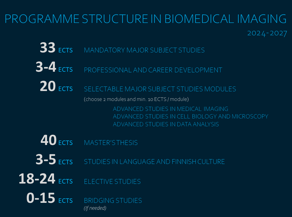
For the programme course structure, see:
Biomedical Imaging course structure – ÅAU
Biomedical Imaging course structure – UTU
Overview:
The application period for this programme opens annually in January.
The studies begin in late August in the following autumn semester.
See the exact application dates in the study info of ÅAU and UTU.
The MSc Programme in Biomedical Imaging is jointly administered by the cluster of Biochemistry and Cell biology, at the Department of Natural and Health Sciences at Åbo Akademi University and the Faculty of Medicine at the University of Turku. In order to increase the chances of being admitted, applicants are advised to apply to both Universities.
This international programme is intended for students with a BSc degree in fields such as biochemistry, bioinformatics, biology, biomedical sciences, chemistry, computational biology, engineering, medical sciences, pharmacology or physics.
Applications are reviewed by the admission committee, which evaluates applicants based on their academic performance, academic background, and relevant skills.
Applying step-by-step:
1. Check both the general and program-specific instruction of both universities on how to apply
Master programmes | Åbo Akademi University
Master programmes | University of Turku
Programme-specific admission criteria | Åbo Akademi University
Programme-specific admission critera | University of Turku
2. Complete language tests if applicable
Language Requirements | Åbo Akademi University
Language Requirements | University of Turku
3. Submit application through study info portal
Opintopolku | Åbo Akademi University
Opintopolku | University of Turku
Possibilities after graduation
Students choose study modules within the program, along with elective courses that align with their individual career goals.
After completing their studies, students can work in various fields both in Finland and abroad.
Examples of career paths our graduates have pursued:
- continue as postgraduate students to pursue a career as a scientist,
- work in core facility management,
- work in science administration nationally or internationally,
- work in hospital research laboratories,
- work in industry and industrial research, or
- work in imaging network or project management.
For more information on studying in Turku, visit the websites of the universities for admitted students.
Information for admitted students | Åbo Akademi University
Information for admitted students | University of Turku
Not available at the moment.
Student experiences
Toiba, India
(started 2018)"Now when studying in the Biomedical Imaging program, I am even more convinced that this is the right path for me."
Imran, Pakistan
(started 2015)"BIMA challenged me to step out of my comfort zone by and helped me to think outside the box. Without this program, I wouldn’t be living my dream of doing a doctorate in Neurology."
Leon, Estonia
(started 2016)"BIMA is an exciting opportunity to go beyond the familiar limits of your field and be at the forefront of the modern scientific research."
Hanna, Finland
(started 2017)
"Earlier I have studied Biotechnology. From the BIMA Program, I get the crucial expertise to my career and in addition, I am able to see the invisible."
Dado,Bosnia and Herzegovina
(started 2018)"It's been a wonderful experience and I love being in Turku. Just go for it and don’t be afraid of the unknown."
Sadaf, Iran
(started 2019)"Even though we have different academic backgrounds, I have decided to follow my sister, who also graduated from Biomedical Imaging few years ago. This proved to be a great decision, and I have no regrets making this life choice”